
Intra-Cerebellar Mouse Functional Connectivity echoes Human Functional Connectivity
Date:
Abstract
Introduction
The cerebellum, traditionally linked to motor control, is now recognized for its role in cognitive and affective processes, with growing evidence of its involvement in pathologies such as autism and schizophrenia1. In humans, neuroimaging studies have characterized cerebellar functional connectivity and organizational gradients2, but analogous investigations in mice remain scarce. Here, we bridge this gap by providing an fMRI protocol optimized for mouse cerebellar imaging, validating connectivity matrices, and deriving functional gradients to enable cross-species comparisons. Our findings reveal conserved connectivity patterns between mice and humans, validating the mouse cerebellar fMRI and functional connectivity methods for cross-species comparisons.
Methods
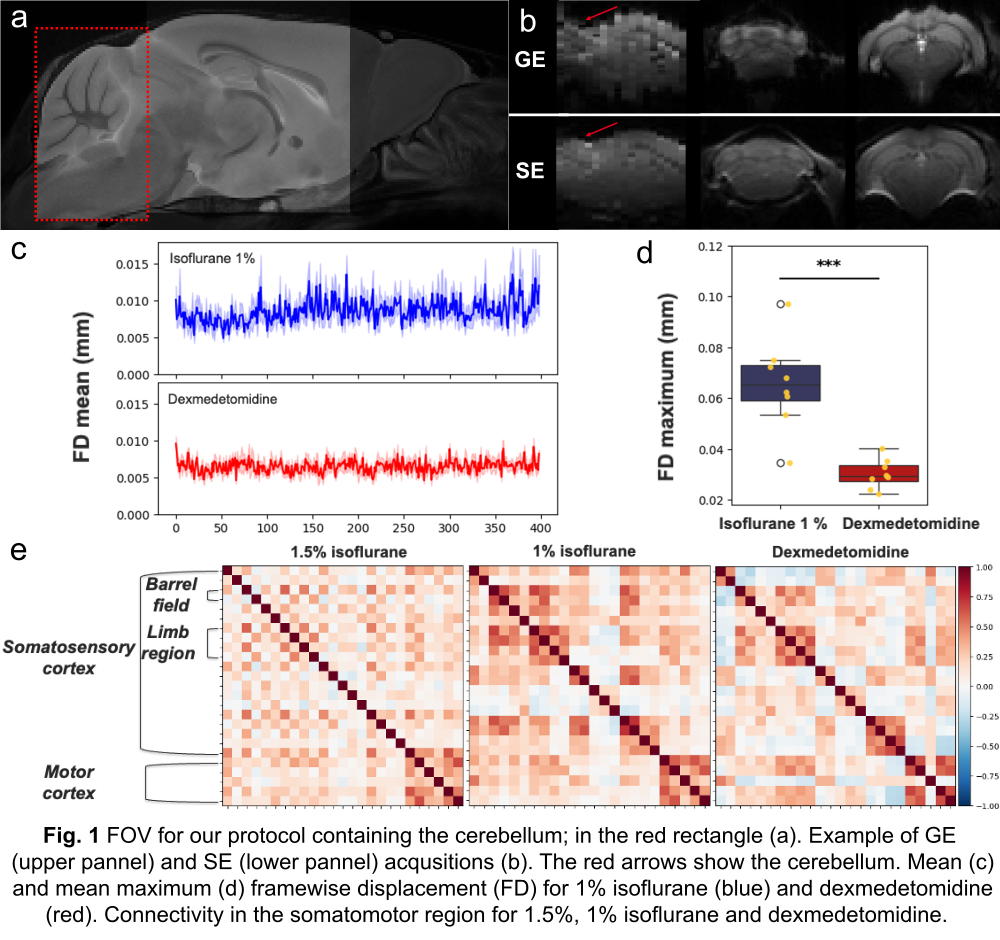
To enable precise functional connectivity studies in mice while maximizing the field of view (Figure 1a), we optimized the fMRI protocol using 17 male C57BL/6J mice (2–4 months old) on an 11.7T Bruker scanner equipped with a 1H-cryoprobe. First, we compared spin-echo (SE) and gradient-echo (GE) echo-planar imaging (EPI). We found that SE had a comparable temporal signal-to-noise ratio (tSNR) to GE but fewer inhomogeneities in the cerebellum (Figure 1b). Next, we evaluated anesthesia protocols: isoflurane (1% or 1.5%) and dexmedetomidine. We observed fewer cerebellar motion artifacts (Figure 1c, d) and stronger connectivity with dexmedetomidine (Figure 1e). The chosen sedation protocol used dexmedetomidine (bolus: 0.025 mg/kg; subcutaneous perfusion: 0.1 mg/kg/h) after isoflurane induction (3%). Isoflurane was halted at least 15 min before scanning (ventilation: air/oxygen 1.5:0.3, 1 L/min). Functional data were acquired using SE-EPI (TR = 1.5 s, TE = 20 ms, 400 volumes, 0.18 × 0.18 × 0.5 mm³ resolution, FOV = 12 × 12 × 9 mm³). Anatomical images were acquired with a Rapid Acquisition with Relaxation Enhancement (RARE) sequence (0.05 × 0.05 × 0.5 mm³ resolution). Besides pilot experiments to set up the protocol, we acquired resting-state fMRI data in 10 wild-type C57BL/6J mice (126.5 ± 0.5 days; 4 females, 6 males). Data were preprocessed using RABIES3, which we adapted for cerebellar-focused analysis. Pearson’s correlation matrices were computed for intra-cerebellar connectivity and cerebellar-cortical connectivity using the DSURQE atlas. We derived 10 functional gradients using cosine similarity, PCA, and Procrustes alignment (BrainSpace4), and the mean gradients were averaged across the group. For cross-species comparisons, we analyzed the HCP Young Adult dataset (n=85). Functional data were acquired using GE-EPI with the following parameters: TR = 1000 ms, TE = 22.2 ms, 1.6 mm isotropic resolution, 85 slices, multiband factor = 5. Data were preprocessed with the HCP Minimal Preprocessing Pipeline5 and Pearson’s correlation matrices were computed with Nilearn. 10 gradients were derived using BrainSpace (PCA, cosine similarity, Procrustes alignment) for intra-cerebellar connectivity, averaged across the group.
Results
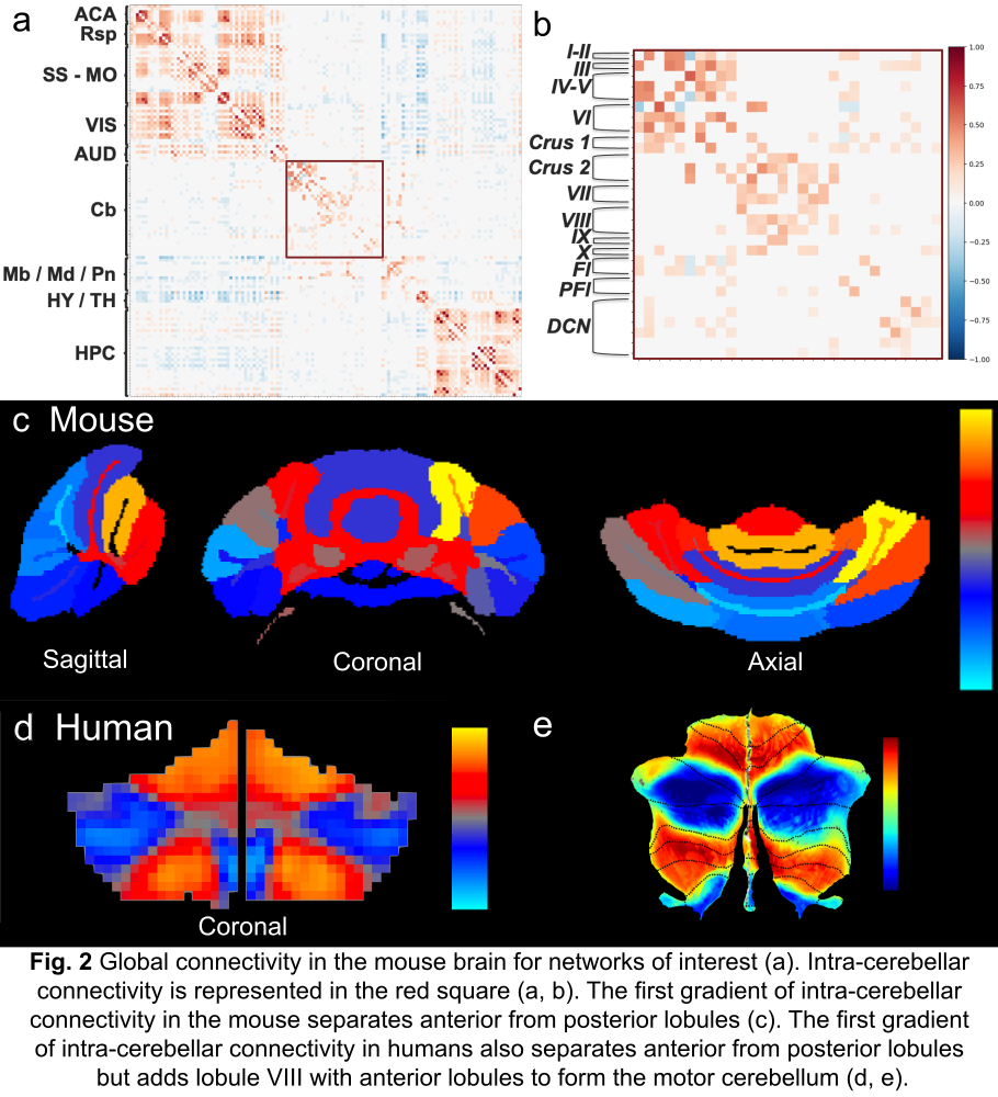
The optimized protocol revealed known functional networks in mice, including the default-mode network (DMN), somatomotor, and visual networks (Figure 2a). We found that cerebellar connectivity towards the rest of the brain was weak for most of the larger brain structures in our FOV. The intracerebellar correlation matrix, in contrast, revealed connectivity patterns between lobules that mirrored human findings (Figure 2b) and displayed a separation between motor and non-motor lobules. The first gradient also reflected this dissociation (Figure 2c), aligning with the human first gradient (Figure 2d, e), showing the motor lobules in red-yellow and the cognitive ones in blue. However, lobule VIII, typically motor-related, clustered with cognitive lobules in both the mouse gradient and the matrix. Furthermore, the separation between motor and non-motor lobules was less distinct in mice. Notably, Crus I showed intermediate connectivity, correlating more with motor lobules than expected. These discrepancies may come from voxel resolution limitations, as our protocol involved a tradeoff between lobule-level resolution and whole-brain coverage.
Conclusions
Our study presents an optimized SE-fMRI protocol for mouse cerebellar imaging, enabling robust functional connectivity analysis. By validating SE-EPI and dexmedetomidine anesthesia, we overcame key technical challenges (motion artifacts and inhomogeneity artefacts) to reveal conserved connectivity patterns between mice and humans. The intra-cerebellar correlation matrix and functional gradients mirrored human findings. Even so, Crus 1 and lobule VIII showed species-specific connectivity, likely due to voxel resolution tradeoffs. The conserved gradients suggest that mouse models can inform human cerebellar dysfunction in pathology. Together, our findings advance preclinical research and pave the way for targeted cerebellar interventions.
References
- Hoppenbrouwers SS, Schutter DJLG, Fitzgerald PB, Chen R, Daskalakis ZJ. The role of the cerebellum in the pathophysiology and treatment of neuropsychiatric disorders: A review. Brain Research Reviews. 2008;59(1):185-200. doi:10.1016/j.brainresrev.2008.07.005
- Guell X, Schmahmann JD, Gabrieli JD, Ghosh SS. Functional gradients of the cerebellum. eLife. 2018;7. doi:10.7554/elife.36652
- Desrosiers-Grégoire G, Devenyi GA, Grandjean J, Chakravarty MM. A standardized image processing and data quality platform for rodent fMRI. Nat Commun. 2024;15(1). doi:10.1038/s41467-024-50826-8
- Vos de Wael R, Benkarim O, Paquola C, et al. BrainSpace: a toolbox for the analysis of macroscale gradients in neuroimaging and connectomics datasets. Commun Biol. 2020;3(1). doi:10.1038/s42003-020-0794-7
- Glasser MF, Sotiropoulos SN, Wilson JA, et al. The minimal preprocessing pipelines for the Human Connectome Project. NeuroImage. 2013;80:105-124. doi:10.1016/j.neuroimage.2013.04.127